Kiran Turaka
Associated Retina Consultants Ltd., USA
Title: Vitreoretinal lymphoma and intravitreal chemotherapy: Ocular and systemic outcomes
Biography
Biography: Kiran Turaka
Abstract
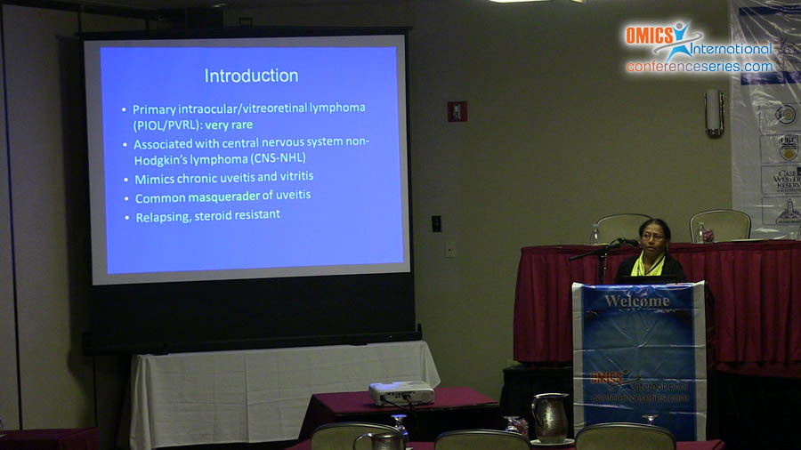
Purpose: Primary intraocular lymphoma is a rare eye malignancy accounting for <1% of all non-Hodgkin’s lymphoma (NHL). Secondary intraocular lymphoma is associated with central nervous system (CNS) NHL and is associated with poor visual and clinical outcomes. The purpose of the study is to repot the clinical, ocular image study findings in patients (pts) with vitreoretinal lymphoma (VRL) and outcomes of treatment with intravitreal chemotherapy. Methods: A retrospective chart review of pts presenting to retina clinic with non-responsive uveitis and vitritis between Jan 2007 to Feb 2012 was performed. Data recorded included demographics, systemic lymphoma status & treatment, ocular symptoms & clinical findings, optical coherence tomography (OCT), fluorescein angiography (FA) & immunocytological findings, treatment methods (intravitreal methotrexate 300 micrograms/0.05 ml, 1000 mcg of rituximab in 0.1 cc) and response. Ocular and systemic lymphoma outcomes at last follow-up visit were noted. Results: Three Caucasian pts (1 female and 2 male) with bilateral vitritis (6 eyes) and CNS- NHL were identified. Mean age of pts was 64.7 years (range 53-80). Visual acuity was better than 20/40 in 4 eyes and ≤ 20/200 in 2 eyes at presentation. Iritis and uveitis were seen in 2 (33.3%) eyes and vitritis in all 6 eyes (100%). Yellowish-white subretinal infiltrates in peripapillary & macular region were present in 4 eyes (66.7%). Mean central foveal thickness on OCT was 270 (range 215-371) μm. Cystoid macular edema was present in 3 eyes (50%), subretinal fluid in 2 (33.3%) & RPE irregularities in 4 (66.7%) eyes. Most common FA findings were macular edema in 3 eyes and perivascular leakage in 1 eye. Immunocytological analysis revealed elevated levels of IL-6 (26.7 pg/ml), IL-10 (12783.5 pg/ml) and IgH gene rearrangement suggestive of lymphoma. All 6 eyes were treated with intravitreal methotrexate (mean 9.7, range 2-15). Mean duration between first intravitreal methotrexate injections to the treatment response was 3.7 weeks (range 3-7). Two pts developed keratitis secondary to methotrexate toxicity. Of which one was treated with intravitreal rituximab (2 injections in right eye and 4 in left eye) for persistent vitritis. First pt developed keratitis after 6 months of six methotrexate injections (monthly) and second patient developed after 5 weeks of seven injections (biweekly & weekly). None of the pts developed rituximab related side effects. Mean duration of follow-up was 15 months (range 4-33). At last follow-up, VRL was persistent in 4 eyes and resolved in 2 eyes. Systemic disease was in remission in 2 pts. Visual acuity was better than 20/40 in 5 eyes and ≤ 20/200 in 1 eye. One patient CNS-NHL died 6 months after treatment of VRL due to CNS relapse. He was treated with 15 intravitreal injections of methotrexate for VRL and also was treated with systemic chemotherapy and radiotherapy for CNS-NHL. Conclusions: Intravitreal methotrexate and rituximab were effective in controlling the VRL with less local side effects. Primary CNS-NHL is associated with poor survival among these pts though VRL is well controlled with localized chemotherapy.